Your Vision
In Expert Hands

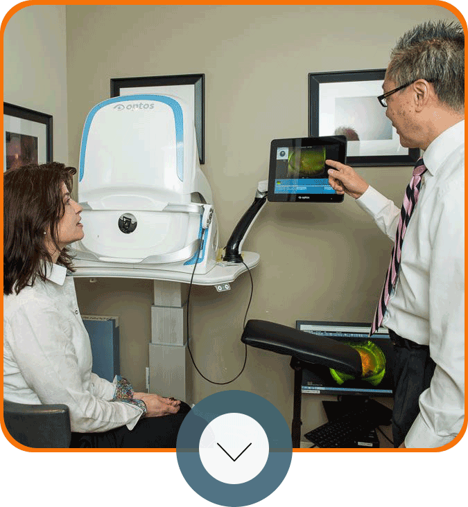
Patient receiving advanced retinal eye care
4.9 (597)
4.9 (419)
4.9 (651)
4.8 (455)

Our Retina Specialists

Gregory Haffner, M.D.

Gregory Haffner, M.D.

Retina Specialist and Vitreoretinal Surgeon

Marvin Sears Award

Excellence in Clinical Ophthalmology, Yale-New Haven Medical Center

Board Certified

American Board of Ophthalmology

Fellow

American Academy of Ophthalmology

Member

American Society of Retina Specialists

Clinical Trials Investigator

DRCR Retina Network and industry-sponsored retina trials
John Huang, M.D., MBA

John Huang, M.D., MBA

Vitreoretinal Surgeon and Uveitis Specialist

Board Certified

American Board of Ophthalmology, since 2005

Founder and President

Connecticut Uveitis Foundation

Committee Member

World Health Organization Global Burden of Disease

Clinical Associate Professor

Yale School of Medicine

Residency Program Director

Yale University Department of Ophthalmology, 2007-2016

Director, Uveitis Service

Yale University Department of Ophthalmology, 2007-2016

Co-Director, Vitreoretinal Service

Yale University Department of Ophthalmology, 2007-2016

Chief Resident

Manhattan Eye, Ear, and Throat Hospital, 2002-2003

Certified Physician Executive

American Association for Physician Leadership

Textbook Co-Author

Ocular Inflammatory Disease and Uveitis Manual, Lippincott Williams & Wilkins

Textbook Co-Author

Ophthalmology Oxford Handbook, Oxford University Press

Member

AAO, ASRS, AUS, CSEP
Erol Verter, M.D.

Erol Verter, M.D.

Retina Specialist and Vitreoretinal Surgeon

Board Certified

American Board of Ophthalmology

Fellow

American Academy of Ophthalmology

Member

American Society of Retina Specialists

Clinical Instructor

Yale School of Medicine, Department of Ophthalmology

Research Grant

Yale Diabetes Research Center, 2018-2020
John Siano, DO

John Siano, DO

Vitreoretinal Surgeon and Retina Specialist

Board Certified

American Board of Ophthalmology

Chief Resident

Nassau University Medical Center

Graduated with Honors

New York College of Medicine

Member

American Academy of Ophthalmology

Member

American Society of Retina Specialists

Member

Connecticut Society of Eye Physicians
Illustration of intraocular lens types used in cataract surgery

Specialized Care,
Personal Attention

For 30 years, New England Retina Associates has been a trusted referral center for optometrists, ophthalmologists, and patients across Connecticut. With four fellowship-trained vitreoretinal surgeons and four locations across Fairfield and New Haven Counties, NERA provides the full spectrum of medical and surgical retina care. From routine monitoring to complex surgery, every patient receives individualized treatment guided by decades of specialized experience. That singular focus means every member of our team, every piece of equipment, and every clinical decision is centered on one thing: the health of your retina. With more than 2,000 five-star reviews and 30 years of care, patients and referring doctors throughout Connecticut trust NERA for conditions that demand true retinal expertise.

Insurance Partners
Aetna insurance logo Cigna insurance logo Medicaid logo Anthem insurance logo Aetna Medicare logo Medicare logo Beneficiaries insurance logo Harvard Pilgrim Health Care logo Empire Blue Cross Blue Shield logo GHI insurance logo Connecticare insurance logo Connecticare VIP logo United Healthcare logo Aetna insurance logo Cigna insurance logo Medicaid logo Anthem insurance logo Aetna Medicare logo Medicare logo Beneficiaries insurance logo Harvard Pilgrim Health Care logo Empire Blue Cross Blue Shield logo GHI insurance logo Connecticare insurance logo Connecticare VIP logo United Healthcare logo

Treatments & Procedures

From intravitreal injections to advanced microsurgery, every treatment plan is tailored to your diagnosis and designed to protect your vision long-term.

View All Treatments

Training the Next Generation of Retina Specialists

Few retina practices in the region train future specialists. New England Retina Associates does, through a two-year vitreoretinal surgery fellowship program. That distinction reflects the depth of expertise our physicians bring to every patient visit. When a practice is trusted to teach the most advanced retinal procedures, the patients it serves receive that same level of precision and knowledge.

New England Retina Associates staff group photo

30 Years of Care & Commitment

Google Reviews

Access to Tomorrow's Treatments

NERA is an active clinical trial site, with physicians serving as investigators in studies for macular degeneration, geographic atrophy, diabetic macular edema, and retinal vein occlusion. For patients who qualify, clinical trials can offer access to new treatment options alongside the highest standard of monitoring and follow-up care.

advanced retina care
Smiling patient asking cataract surgery questions

NERA's fellowship-trained retina specialists treat the full spectrum of retinal and vitreous conditions. Whether you have been referred by your eye doctor or are seeking care on your own, every NERA location is equipped to evaluate and treat the following conditions.

Macular & Degenerative Conditions

  • Age-Related Macular Degeneration (Dry AMD) — The most common cause of central vision loss in adults over 50, caused by the gradual breakdown of light-sensitive cells in the macula. NERA physicians monitor dry AMD closely and guide patients through evidence-based management, including nutritional supplementation and regular retinal imaging.
  • Age-Related Macular Degeneration (Wet AMD) — An advanced and more rapidly progressing form in which abnormal blood vessels grow beneath the retina, leaking fluid and damaging central vision. Anti-VEGF injections are the primary treatment for wet AMD and are highly effective when started early.
  • Geographic Atrophy — An advanced stage of dry AMD involving the progressive loss of retinal pigment epithelium and photoreceptor cells. FDA-approved treatments for geographic atrophy are now available, and NERA physicians are actively involved in clinical research for this condition.
  • Macular Hole — A small break in the center of the macula that causes blurred or distorted central vision. Vitrectomy surgery is the primary treatment and achieves high closure rates in appropriately selected patients.
  • Macular Pucker (Epiretinal Membrane) — A thin layer of scar tissue that forms on the surface of the macula, causing central vision to appear distorted or wrinkled. Observation is appropriate for mild cases; vitrectomy is recommended when vision is meaningfully affected.
  • Central Serous Retinopathy (CSR) — A condition in which fluid accumulates beneath the retina, typically affecting younger to middle-aged adults and often associated with stress or corticosteroid use. Many cases resolve on their own; chronic or recurrent cases may benefit from photodynamic therapy or laser treatment.
  • Macular Telangiectasia — A rare condition involving abnormal capillaries near the center of the macula. NERA physicians monitor for progression and provide treatment when neovascular complications develop.

Diabetic & Vascular Conditions

  • Diabetic Retinopathy — Progressive damage to the blood vessels of the retina caused by long-standing diabetes. One of the leading causes of vision loss in working-age adults in the United States; early detection and treatment can substantially reduce the risk of permanent vision impairment.
  • Diabetic Macular Edema (DME) — A complication of diabetic retinopathy in which fluid accumulates in the macula, causing central vision to blur or become distorted. Anti-VEGF injections are effective for many patients with DME, and other treatment options are available when needed.
  • Retinal Vein Occlusion (BRVO / CRVO) — A blockage of the veins that drain blood from the retina, leading to bleeding, swelling, and vision loss. Both branch retinal vein occlusion (BRVO) and central retinal vein occlusion (CRVO) are evaluated and treated at NERA.
  • Retinal Artery Occlusion — A sudden blockage of the artery supplying the retina, often presenting as painless, rapid vision loss in one eye. Retinal artery occlusion is a retinal emergency requiring prompt evaluation. NERA accepts urgent and emergent referrals.

Structural & Emergency Conditions

  • Retinal Detachment — A medical emergency in which the retina separates from the underlying tissue. Warning signs include a sudden increase in floaters, flashing lights, or a dark curtain across your vision. NERA handles retinal emergencies and accepts urgent referrals from emergency rooms and eye care providers.
  • Retinal Tears — A tear in the retina that can progress to a full detachment if left untreated. Laser retinopexy or cryoretinopexy can seal a tear and prevent further complications in most cases.
  • Horseshoe Tear — A specific type of retinal tear caused by vitreous traction on the retina, characterized by a horseshoe shape. Prompt treatment is recommended to reduce the risk of detachment.
  • Floaters and Flashes — Most floaters are benign and related to normal changes in the vitreous gel. However, a sudden onset of new floaters, light flashes, or a shadow in your vision warrants prompt evaluation to rule out a retinal tear or detachment.
  • Complications After Cataract Surgery — Retinal complications following cataract surgery — including cystoid macular edema, retinal detachment, vitreous hemorrhage, or a dislocated intraocular lens — require subspecialty retinal care. NERA physicians evaluate and manage a full range of post-cataract retinal complications and can perform scleral-fixated IOL implantation when needed.

Inflammatory & Other Conditions

  • Uveitis (Posterior / Panuveitis) — Inflammation involving the retina, vitreous, or choroid that can lead to serious vision loss if inadequately treated. NERA's Dr. John Huang holds dedicated fellowship training in uveitis and ocular immunology from Harvard University and coordinates care with rheumatology and other specialists when systemic disease is involved.
  • Intraocular Tumors — Abnormal growths within the eye that require specialized imaging and evaluation. NERA physicians assess lesion characteristics carefully and coordinate with ocular oncology specialists when appropriate.
  • Central Serous Chorioretinopathy — Fluid accumulation between the retina and retinal pigment epithelium, most often in middle-aged patients. NERA physicians manage both acute and chronic presentations with monitoring, photodynamic therapy, or laser treatment as indicated.

NERA provides a comprehensive range of medical and surgical retinal treatments. Every treatment plan is determined by your physician based on your specific diagnosis, the severity of your condition, your treatment history, and your overall health. Results vary by individual; your NERA physician will discuss the most appropriate approach for your situation.

Injection Therapies

Intravitreal (in-the-eye) injections are the most widely used treatment in retina medicine today. Anti-VEGF medications reduce abnormal blood vessel growth and fluid leakage in the retina and are the cornerstone of treatment for wet AMD, diabetic macular edema, and retinal vein occlusions. NERA physicians use all leading anti-VEGF agents, including:

  • Aflibercept (Eylea)
  • Faricimab (Vabysmo)
  • Bevacizumab (Avastin)
  • Ranibizumab (Lucentis)

Your doctor will recommend the most appropriate medication based on your condition and how your retina responds over time. In some cases, steroid injections — including intravitreal triamcinolone or sustained-release implants — are used to reduce inflammation and fluid in conditions such as uveitis, macular edema, and certain retinal vein occlusions.

Laser Treatments

  • Focal and Grid Laser — Targets specific areas of fluid leakage in the macula, used for select cases of diabetic macular edema and other macular conditions.
  • Panretinal Photocoagulation (PRP) — A broad laser treatment applied to the peripheral retina to reduce abnormal blood vessel growth in proliferative diabetic retinopathy and other advanced retinal diseases.
  • Laser Retinopexy — Creates small laser burns around a retinal tear to form a seal, preventing fluid from passing beneath the retina and progressing to detachment.
  • Photodynamic Therapy (PDT) — A light-activated treatment in which a photosensitizing medication is administered intravenously and then activated by a low-energy laser. Used for certain types of wet AMD and chronic central serous retinopathy.
  • Transscleral Diode Laser (Non-Thermal) — A specialized, non-thermal infrared laser approach available at NERA for specific retinal conditions where conventional thermal laser is not appropriate.

Surgical Procedures

  • Pars Plana Vitrectomy — A microsurgical procedure in which the vitreous gel is carefully removed from inside the eye and replaced with a clear solution or gas bubble. Vitrectomy is the primary surgical treatment for retinal detachments, macular holes, epiretinal membranes, diabetic vitreous hemorrhage, and severe vision-impairing floaters. It is also used to manage complex retinal complications following cataract surgery.
  • Scleral Buckle — A surgical repair technique for retinal detachment in which a soft silicone band is sutured around the outside of the eye to gently push the eye wall inward and support the detached retina. Often used in combination with vitrectomy or as a standalone procedure for appropriate cases.
  • Pneumatic Retinopexy — An office-based procedure for select retinal detachments involving injection of a small gas bubble into the eye, followed by specific head positioning to allow the bubble to press against and reattach the retina. No operating room visit is required for this procedure.
  • Cryoretinopexy — Uses a precisely controlled application of extreme cold to create a scar that seals retinal tears and prevents fluid from passing beneath the retina. Often used in combination with scleral buckling or pneumatic retinopexy.
  • Scleral-Fixated IOL Implantation — A specialized surgical technique to anchor an intraocular lens to the scleral wall of the eye when the natural lens capsule is absent or damaged. Most often needed after complicated cataract surgery or trauma in which a standard IOL cannot be supported.

An accurate diagnosis is the foundation of effective retinal care. NERA uses advanced imaging technology to detect retinal disease at its earliest stages, monitor changes over time, and guide treatment decisions with precision. Most of these tests are quick, non-invasive, and completed in the office during your visit — no hospital trip required.

  • Optical Coherence Tomography (OCT) — The most widely used retinal imaging tool in modern practice, OCT creates detailed, cross-sectional images of the retina's layers at near-microscopic resolution, revealing fluid accumulation, structural changes, and early signs of disease. The scan is completely non-invasive, requires no contact with the eye, and takes only seconds. OCT imaging is used at virtually every NERA visit and is essential for monitoring treatment response.
  • OCT Angiography (OCTA) — An advanced extension of OCT that maps blood vessel flow in the retina and choroid without requiring any dye injection. OCT angiography is particularly valuable for monitoring wet AMD, diabetic eye disease, retinal vein occlusions, and other vascular conditions. Like standard OCT, it is non-invasive and takes only minutes.
  • Wide-Field Retinal Imaging — Captures a panoramic photograph documenting a far greater area of the retinal surface than traditional fundus cameras. Wide-field imaging is especially useful for detecting peripheral retinal disease, staging diabetic retinopathy, identifying retinal tears, and creating a baseline record of retinal health. The test is brief, non-invasive, and involves only a bright flash of light.
  • Fluorescein Angiography (FA) — A dye-based imaging test in which fluorescein — a bright yellow dye — is administered through a small intravenous injection and then photographed as it circulates through the retinal blood vessels. Fluorescein angiography reveals leaking vessels, areas of blockage, and abnormal vascular growth with a level of detail not possible with standard photography. Skin and urine may appear temporarily yellowish after the test; this resolves on its own within hours.
  • Indocyanine Green Angiography (ICG) — Similar in principle to fluorescein angiography but uses a different infrared-absorbing dye to visualize the deeper choroidal blood vessels beneath the retina. ICG angiography provides critical information in conditions such as central serous retinopathy, certain presentations of AMD, and polypoidal choroidal vasculopathy, where the standard FA alone may not provide a complete picture.
  • Ophthalmic Ultrasound (B-Scan) — Uses high-frequency sound waves to image the interior of the eye, particularly when the view through the front of the eye is blocked by a dense cataract, vitreous hemorrhage, or other opacity. Ultrasound is also used to evaluate the vitreous and peripheral retina and to assess suspected intraocular tumors. A small probe is placed gently on the closed eyelid; the test is entirely painless.
  • Fundus Autofluorescence (FAF) — A specialized imaging technique that measures the natural fluorescence of the retinal pigment epithelium, revealing areas of cell damage or dysfunction. FAF is particularly useful for monitoring geographic atrophy, inherited retinal dystrophies, and other conditions affecting the outer retina and RPE.
  • Genetic Testing — Identifies genetic variants associated with inherited retinal diseases such as Stargardt disease, Best disease, retinitis pigmentosa, and choroideremia. Genetic test results can confirm a diagnosis, guide prognosis, assist with family planning, and identify patients who may be candidates for gene-based therapies currently in development. Testing typically involves a simple blood draw or cheek swab; results are usually available within a few weeks.

New England Retina Associates has provided subspecialty retinal care to Connecticut patients since 1995. The practice is led by four fellowship-trained vitreoretinal surgeons with training backgrounds at Yale, Harvard, and Johns Hopkins — each board-certified by the American Board of Ophthalmology. With more than 2,000 five-star patient reviews across four Connecticut locations, NERA serves as a major referral center for optometrists and ophthalmologists throughout Fairfield County, New Haven County, and neighboring New York. The practice also operates an on-site two-year vitreoretinal surgery fellowship program, reflecting its role as both a clinical and academic institution.

Gregory Haffner, M.D. — Partner, Vitreoretinal Surgeon

Dr. Haffner has been at NERA for more than 20 years and serves as a partner in the practice. He completed his ophthalmology residency at Yale University, where he received the Marvin Sears Award for Excellence in Clinical Ophthalmology, before completing his vitreoretinal surgery fellowship at NERA itself. Dr. Haffner is an active clinical trial investigator in age-related macular degeneration, diabetic retinopathy, and retinal vein occlusions, and has been recognized nationally as one of America's Top Ophthalmologists by the Consumers' Research Council of America.

John Huang, M.D., MBA, C.P.E. — Vitreoretinal Surgeon, Uveitis Specialist

Dr. Huang holds dual fellowship training from Harvard University — where he trained in uveitis and ocular immunology at Massachusetts Eye and Ear Infirmary — and from Johns Hopkins, where he completed his vitreoretinal surgery fellowship at the Wilmer Eye Institute. This combination is held by very few retina specialists in the region. Dr. Huang served on the Yale faculty for seven years as Associate Professor of Ophthalmology, Program Director of the residency, and Director of the Uveitis Service. He is the founder of the Connecticut Uveitis Foundation, a textbook author, and a member of the Committee for Global Burden of Disease at the World Health Organization.

Erol Verter, M.D. — Retina Specialist, Vitreoretinal Surgeon

Dr. Verter's training spans Harvard Medical School (four years of ophthalmology research at Massachusetts General Hospital), Boston University School of Medicine, and Yale-New Haven Hospital, where he completed his ophthalmology residency before returning to NERA for his vitreoretinal surgery fellowship. He serves as a Clinical Instructor of Ophthalmology at Yale School of Medicine and as Chair of Ophthalmology at St. Vincent's Medical Center, and holds hospital appointments at Yale New Haven, Bridgeport, Stamford, Greenwich, and Saint Vincent hospitals. Dr. Verter conducted Yale Diabetes Research Center-funded research on contrast sensitivity as an early detection tool for diabetic retinopathy, and volunteers at the Dana Eye Clinic at Yale New Haven Health, providing care for uninsured patients throughout the community.

John Siano, D.O. — Retina Specialist, Vitreoretinal Surgeon

Dr. Siano completed his ophthalmology residency at Nassau University Medical Center, where he served as Chief Resident, before completing his vitreoretinal surgery fellowship at NERA. His clinical focus includes macular degeneration, geographic atrophy, diabetic retinopathy, diabetic macular edema, retinal vein occlusion, macular hole, retinal detachment, and retinal complications following cataract surgery. Dr. Siano is actively involved in clinical trials for AMD and geographic atrophy, giving eligible patients access to emerging therapies before they are widely available. He is currently accepting new patients at all NERA locations.

Our Locations


2200 Whitney Avenue, Suite 300
Hamden, CT 06518


Get Directions
Map for Hamden


999 Silver Lane, 2B
Trumbull, CT 06611


Get Directions
Map for Trumbull


162 Kings Highway North
Westport, CT 06880


Get Directions
Map for Westport


1445 East Putnam Avenue
Old Greenwich, CT 06870


Get Directions
Map for Old Greenwich
Map for Hamden
Map for Trumbull
Map for Westport
Map for Old Greenwich